


An anomaly scan is an important prenatal screening and is a crucial step as it determines the health of the developing fetus and the expectant mother. To get the benefits of this ultrasound examination, the parents-to-be must be ready mentally and physically and know what to expect. Here, you will learn more about why an anomaly scan is done, what it helps determine and more.

An anomaly scan, also called a 20-week pregnancy scan, is an ultrasound performed to assess the fetus’s physical development and identify any structural abnormalities or potential concerns.
The anomaly scan is typically performed between 18 and 22 weeks of pregnancy, with most healthcare providers recommending it around 20 weeks. It can be performed between weeks 18 and 21 of pregnancy. It also helps to detect congenital disorders and anatomical abnormalities. The scan provides 2D ultrasound imaging, though 3D or 4D images may be used when needed. During the procedure, the sonographer takes detailed measurements to evaluate whether the baby's growth and development are appropriate for the gestational age.
A 20-week pregnancy scan is safe because it does not use X-rays or CT scans; instead, it uses sound waves. Your healthcare provider may recommend a 20-week scan, but undergoing the scan is optional, though it is preferable to get it done so that you know the fetus is healthy.
This ultrasound scan measures your baby's body parts and organs to ensure healthy, normal development throughout pregnancy. The scan also looks for structural issues with organs and specific congenital abnormalities. Some of the key areas examined include:
The sonographer measures head size and checks the brain's internal structures to ensure proper development in accordance with standards. They also scan the entire spine to confirm that the vertebrae are aligned and that the protective skin covers the entire length of the spinal cord. This scan is required to rule out spina bifida (a congenital neural tube defect) or any fluid buildup in the brain.
During an anomaly scan, the sonographer closely examines the function of the heart and the major blood vessels connected to it. They monitor the heart rhythm and note whether the valves open and close correctly. If any congenital heart defects are present, they are detected at this stage.
The sonographer checks the lungs for any unusual cysts or fluids. They also check if the diaphragm is intact and properly separates the chest from the abdomen.
The anomaly scan confirms whether the abdominal wall is fully formed and completely covers all the internal organs. The sonographer checks if the baby is actively swallowing amniotic fluid. The position and appearance of the bowels are also checked.
The scan checks for the presence of both kidneys and for normal urine production. They also check the baby’s bladder.
The sonographer measures the lengths of the hands and legs to match the standards for expected gestational growth. They also examine the hands and feet to rule out conditions such as clubfoot.
The sonographer looks at the baby’s face from multiple angles to assess the structure of the nose, lips, and palate. They also check whether the eyes are positioned correctly and whether the spacing between them is appropriate.
The anomaly scan checks the exact position of the placenta to confirm that it is not blocking the cervix. It also measures the volume of the amniotic fluid to ensure that the baby has enough space to move and grow safely. The sonographer also checks the three blood vessels inside the umbilical cord to confirm that nutrients are delivered properly to the baby.
The length of the cervix is measured to ensure that it is closed and long enough to support pregnancy.
During an ultrasound, several images are taken, and the sonographer will draw lines on them. This is a way to measure the size of the limbs and organs. It is then compared with your due date, and if it is within a fortnight of the predicted due date, the fetus is said to be developing properly. The baby's measurements are compared with standard growth parameters for the gestational age to ensure healthy development and identify any potential concerns.

An anomaly scan is used to:
The step-by-step procedure of an anomaly scan is as follows:
You’ll be asked to lie on your back, usually on an examination table, and expose your lower abdomen to give the healthcare provider clear access for the scan.
A warm, clear conductant gel is applied across your belly. The healthcare provider will gently move a handheld transducer over your abdomen, producing real-time images on the monitor using sound waves.
The sonographer checks your baby’s anatomy from head to toe to match structural landmarks. They analyse the heart chambers, brain symmetry, facial features, the spine, and individual internal organs.
The sonographer maps the precise position of your placenta to rule out conditions such as a low-lying placenta. They measure the amniotic fluid volume to ensure your baby is safely cushioned and supported.
Standard physical measurements are noted, specifically the head circumference, abdominal width, and femur (thigh bone) length. These measurements are compared to ensure your baby's weight and growth milestones align with your stage of pregnancy.
A complete, comprehensive anomaly evaluation typically takes between 30 and 45 minutes to finish. The timeline may vary slightly depending on how active your baby is and their position in the womb.
The ultrasound process is completely non-invasive and safe for both mother and child. You might only feel mild, temporary physical discomfort when the specialist needs to apply firmer pressure to capture clear images of a shy or poorly angled baby.
You will receive immediate baseline feedback during imaging, along with printed keepsake photos. A comprehensive diagnostic report will be officially prepared for your primary doctor to review with you.
If your baby is in an awkward position, a simple walk or a repeat scan may be recommended to capture any missed details. If any developmental variations or a low-lying placenta are detected, your doctor may discuss the next steps, ask you to go for specialised counselling, or organise a target fetal echo scan.
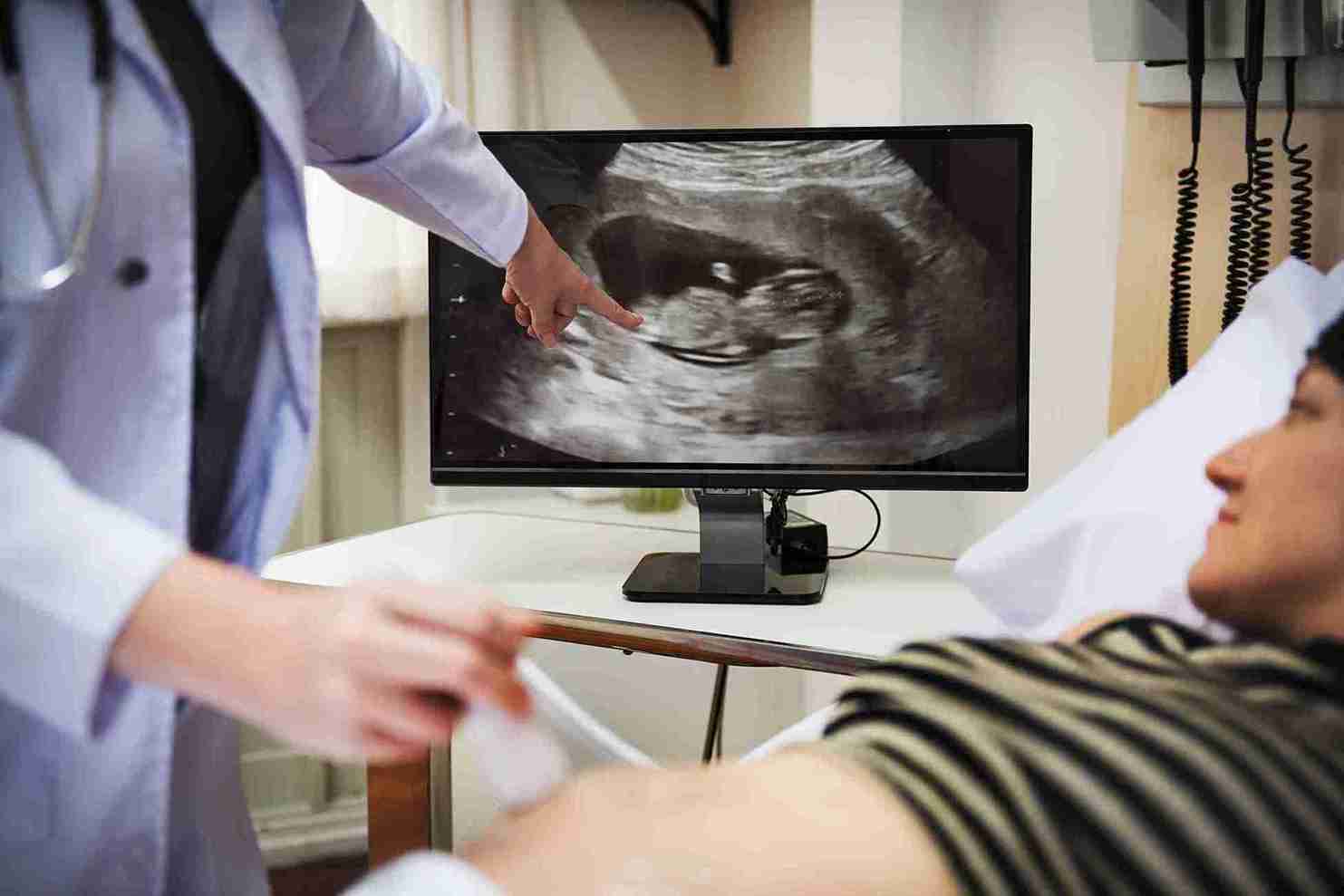
While anomaly scans can detect many structural abnormalities, they cannot identify every condition. While most of the defects can be identified, not all of them can be. Some defects can arise later in pregnancy and may not be detected at 20 weeks. So, an anomaly scan does not give you a 100% guarantee of a healthy baby at birth. However, this does not mean the scan should be dismissed, as it can detect most abnormalities—particularly when performed with advanced ultrasound technology by experienced sonographers. Here are some conditions that may not be detected by the anomaly scan:
If you learn that an anomaly scan has identified a possible abnormality, it can be worrying for expectant parents. Remember: an abnormal finding does not always mean that your baby has a serious health issue. In some cases, the scan may detect something that may turn out to be harmless. In other cases, it may require additional evaluation to understand the significance.
If an abnormality is suspected, your healthcare provider may recommend additional tests or follow-up assessments. It may be a repeat ultrasound, a fetal echocardiogram, or any diagnostic test. These investigations can give more information and help to confirm or rule out certain conditions.
One of the key advantages of an anomaly scan is that it helps identify any potential risks. Early detection leads to better monitoring, appropriate planning, and, if required, treatment before or immediately after birth. It gives expectant parents time to understand the diagnosis, discuss available options, and make decisions based on their doctor's recommendations.
If you find further testing to be overwhelming, remember that many pregnancies with abnormal scan findings have ended up with positive results. Your doctor can support you throughout the process and answer any questions or concerns as they arise.
There is no preparation needed for an anomaly scan appointment. The scan itself will take about 45 minutes, and if you see your doctor with the results, that can add about 30 minutes more. So the appointment may last about 75 minutes in all. The anomaly scan price can start at Rs 1000 and depends on where you get it done. That is the only preparation you need for the scan. But the only thing needed is to get the baby in position to get clear images.
Here are a few tips to get the baby moving, as sometimes babies make it difficult to get clear images. Here are a few tips to help your baby get into a better position.
Further tests may be recommended if an anatomy scan of the pregnancy shows something is wrong. You can discuss it with your healthcare provider and decide if you want to go ahead with it.

In addition to the anomaly scans, there are a few other scans that can help you identify if the baby is developing as expected.
The NT scan is performed between 11 and 14 weeks of pregnancy, and it measures the fluid at the back of the baby’s neck. This scan helps identify the risk of chromosomal conditions such as Down Syndrome.
A growth scan is performed in the 3rd trimester to monitor the baby's weight, position, and overall growth and well-being. This helps to ensure that the baby is growing appropriately and is getting enough nutrition and oxygen.
A Doppler scan checks the blood flow between the mother, placenta and the baby. It is normally recommended for any high-risk pregnancies to assess whether the baby is getting enough oxygen and nutrients from the mother.
Fetal echocardiography is an ultrasound which studies the baby’s heart structure and function. It is commonly recommended if there is a family history of heart defects, abnormal scan findings, or other risk factors for congenital heart conditions.
The anomaly scan is a key pregnancy milestone, offering valuable insight into your baby's growth and development around 20 weeks. This safe, non-invasive scan examines major organs and structures to detect potential abnormalities early, enabling timely care when needed. While it cannot guarantee a completely risk-free outcome, it remains an essential tool that provides reassurance and helps ensure a healthy pregnancy journey.


The NT scan is done between 11 and 14 weeks and measures neck fold fluid to assess the risk of chromosomal conditions like Down Syndrome. The anomaly scan, performed around 18–22 weeks, provides a detailed structural evaluation of the baby's brain, heart, spine, organs, limbs, and facial features, and also assesses the placenta, amniotic fluid, and cervix to detect physical malformations and congenital abnormalities.
No, the anomaly scan is completely non-invasive and pain-free. You may feel mild, temporary discomfort if the sonographer applies firmer pressure to get clearer images, especially if the baby is positioned awkwardly. Overall, it involves a gel applied to the belly and a probe gently gliding across the abdomen, making it a comfortable experience for most expectant mothers.
Yes, the anomaly scan is completely safe for both mother and baby. It uses sound waves rather than X-rays or CT rays, making it a non-invasive, risk-free procedure. While it is highly recommended to assess the baby's growth and development and detect potential abnormalities, it remains optional, and the final decision rests with the expectant parents.
No special preparation is needed for the appointment, though wearing comfortable clothing for easy access is advisable. Follow any specific instructions from your healthcare provider regarding hydration or bladder preparation, as requirements can vary depending on individual circumstances and hospital protocol.


 Best Gynecologists in Bellandur Best Gynecologists in Electronic City Best Gynecologists in HRBR LayoutBest Gynecologists in Jayanagar Best Gynecologists in Kanakapura RoadBest Gynecologists in Malleswaram Best Gynecologists in Nagarbhavi Best Gynecologists in Old Airport RoadBest Gynecologists in Sahakarnagar Best Gynecologists in Sarjapur RoadBest Gynecologists in ThanisandraBest Gynecologists in Varthur RoadBest Gynecologists in Whitefield Best Gynecologists in Industrial Area Phase IIBest Gynecologists in OMRBest Gynecologists in T NagarBest Gynecologists in New Industrial TownBest Gynecologists in IndirapuramBest Gynecologists in Golf Course RoadBest Gynecologists in Sector 14Best Gynecologists in Sector 47Best Gynecologists in Banjara HillsBest Gynecologists in GachibowliBest Gynecologists in Hitech CityBest Gynecologists in KompallyBest Gynecologists in Gomti NagarBest Gynecologists in MaladBest Gynecologists in NerulBest Gynecologists in VashiBest Gynecologists in Kailash ColonyBest Gynecologists in PatparganjBest Gynecologists in Punjabi BaghBest Gynecologists in VikaspuriBest Gynecologists in Gaur CityBest Gynecologists in Pari ChowkBest Gynecologists in Sector 51Best Gynecologists in Sector 5 Swastik ViharBest Gynecologists in Kalyani NagarBest Gynecologists in SB RoadBest Gynecologists in Pimple SaudagarBest Pediatricians in BellandurBest Pediatricians in Electronic CityBest Pediatricians in HRBR LayoutBest Pediatricians in JayanagarBest Pediatricians in Kanakapura RoadBest Pediatricians in MalleswaramBest Pediatricians in NagarbhaviBest Pediatricians in Old Airport RoadBest Pediatricians in SahakarnagarBest Pediatricians in Sarjapur RoadBest Pediatricians in ThanisandraBest Pediatricians in Varthur RoadBest Pediatricians in WhitefieldBest Pediatricians in Industrial Area Phase IIBest Pediatricians in OMRBest Pediatricians in T NagarBest Pediatricians in New Industrial TownshipBest Pediatricians in IndirapuramBest Pediatricians in Golf Course RoadBest Pediatricians in Sector 14Best Pediatricians in Sector 47Best Pediatricians in Hitech CityBest Pediatricians in Banjara HillsBest Pediatricians in GachibowliBest Pediatricians in KompallyBest Pediatricians in Gomti NagarBest Pediatricians in MaladBest Pediatricians in NerulBest Pediatricians in VashiBest Pediatricians in Kailash ColonyBest Pediatricians in PatparganjBest Pediatricians in Punjabi BaghBest Pediatricians in VikaspuriBest Pediatricians in Gaur CityBest Pediatricians in Pari ChowkBest Pediatricians in Sector 51Best Pediatricians in Sector 5 Swastik ViharBest Pediatricians in Kalyani NagarBest Pediatricians in SB RoadBest Pediatricians in Pimple Saudagar