



Most women spend the first scan focused entirely on the baby. The heartbeat, the measurements, the tiny flicker of movement on the screen. What often gets less attention, until it becomes a concern, is the other structure that makes all of that possible - the placenta.
It is the organ that grows alongside the baby, delivering oxygen and nutrients, removing waste, and producing hormones that sustain the pregnancy. Where it sits in the uterus, how it is attached, and how it changes across the trimesters all have direct implications for how the pregnancy is managed and how delivery is planned.
For many women, the placenta is only discussed when something about it appears on a scan report—a position that sounds alarming or a term they have never heard before. This guide explains what those terms actually mean, in plain language.
The placenta develops from the same fertilised egg as the baby and attaches to the uterine wall, typically by the end of the first trimester. It grows steadily throughout the pregnancy, reaching about 500 grams and 20 to 25 centimetres in diameter by the time of delivery.
Through the placenta, the baby receives everything it needs and cannot produce on its own. Oxygen, glucose, amino acids, vitamins, and antibodies from the mother all cross the placental barrier. Carbon dioxide and waste products travel back the other way. The placenta also produces progesterone and estrogen, which are essential for maintaining the pregnancy, particularly after the first trimester when the corpus luteum can no longer sustain these hormones alone.

Placenta position during pregnancy is one of the first things assessed on the anomaly scan, typically done around 18 to 20 weeks. The position is described in terms of where the placenta has implanted on the uterine wall.
A posterior placenta is attached to the back wall of the uterus, closest to the mother's spine. This is considered one of the most common and straightforward positions. The baby is typically positioned between the placenta and the mother's abdomen, allowing fetal movements to be noticed more easily and earlier than with other placental positions. Most posterior placentas are associated with uncomplicated pregnancies and do not affect delivery planning unless they are unusually low.
An anterior placenta sits on the front wall of the uterus, between the baby and the mother's abdomen. This is also a common position and, in itself, is not a cause for concern. The placenta in this position acts as a cushion between the baby and the outside, which means fetal movements are often felt later and more faintly than with a posterior placenta.
Many women with an anterior placenta worry when they do not feel kicks as early or as strongly as they expected or as early as other pregnant women describe. This is normal with this placental position and is not a sign that something is wrong. The movements are there, simply dampened by the layer of placental tissue.
A fundal placenta is attached to the top of the uterus. Like posterior and anterior placentas, this is a common and generally uncomplicated position. It sits away from the cervical opening, so it rarely causes problems during delivery.
A lateral placenta attaches to one of the side walls of the uterus and is sometimes described as left lateral or right lateral, based on its exact location. This is a normal variant and does not typically affect the pregnancy or delivery.
Of all the terms that appear on scan reports, low-lying placenta is the one that causes the most anxiety, and it is worth understanding clearly.
A low-lying placenta is one that has implanted in the lower portion of the uterus, close to the cervical opening. When the placenta partially or completely covers the internal cervical os, the opening through which the baby would pass during a vaginal delivery, the condition is called placenta praevia.
A low-lying placenta is identified at the 18-20-week scan and is not a cause for alarm. The uterus grows significantly between 20 weeks and the third trimester, and as it does, the lower segment stretches and the placental attachment point appears to move upward relative to the cervix. This process is sometimes called placental migration, though the placenta itself does not move — the uterus around it grows.
The majority of women who are told they have a low-lying placenta at 20 weeks will have a placenta that has moved well clear of the cervix by 32 to 36 weeks. A follow-up scan, usually at 32 weeks, confirms whether the move has happened.
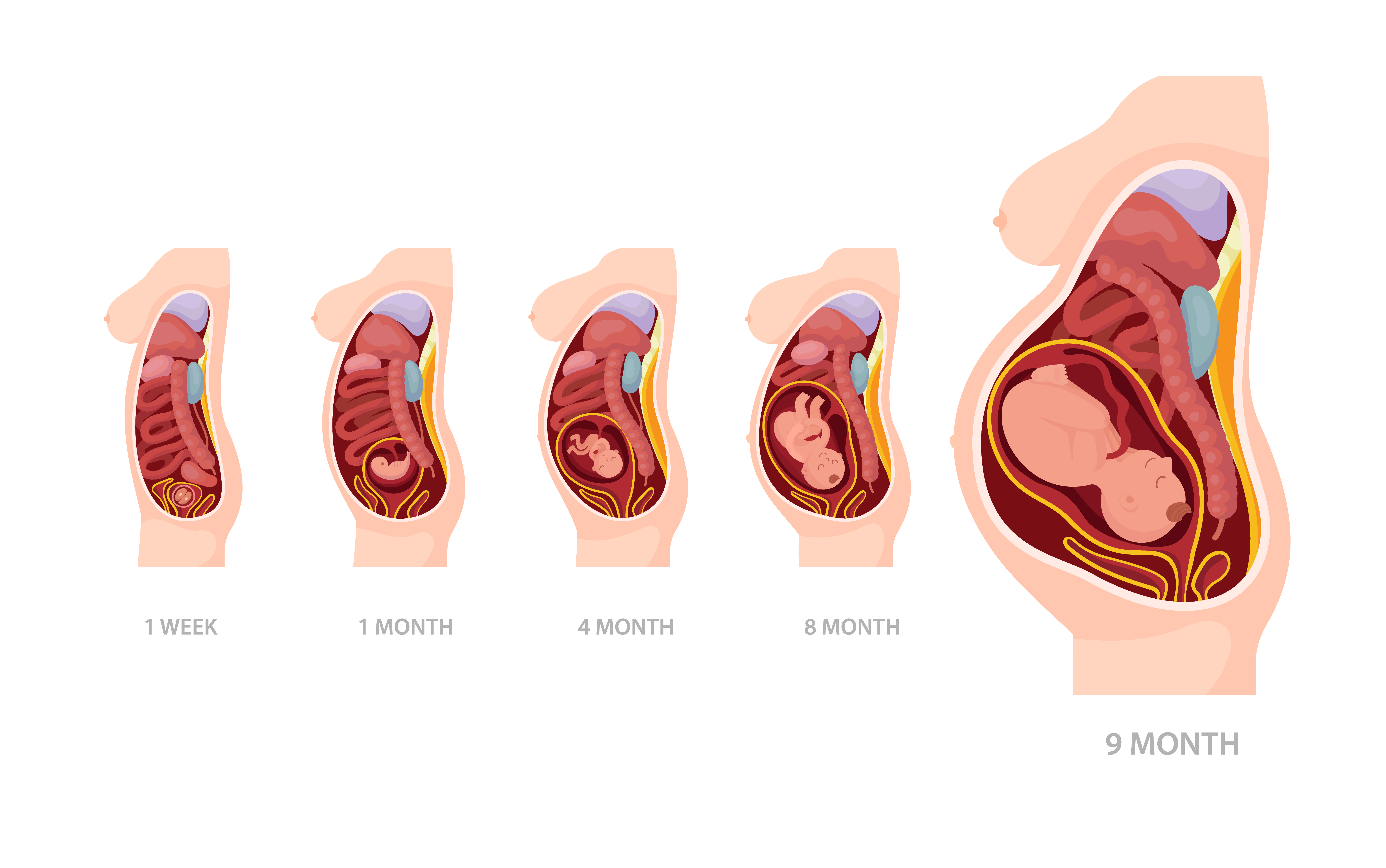
Beyond position, the types of placenta can also be characterised by structural variations that occasionally arise.
In some pregnancies, the placenta develops as two separate lobes of roughly equal size connected by blood vessels and membranes. This is not a dangerous condition in itself, but it does mean the delivery team needs to confirm that both lobes have been delivered completely after birth, as retained placental tissue is a cause of postpartum haemorrhage.
A succenturiate lobe is a smaller additional lobe of placental tissue that develops separately from the main placenta. Like a bilobed placenta, the concern is ensuring complete delivery. The connecting vessels between the main placenta and the accessory lobe can occasionally cross the cervical opening, a condition called vasa praevia, which carries a risk of serious fetal haemorrhage if those vessels rupture.
These terms describe conditions where the placenta attaches too deeply into the uterine wall.
These conditions are more likely to occur in women who have had a previous caesarean section or uterine surgery. They are associated with severe bleeding at delivery and typically require highly specialised obstetric management, including the possibility of hysterectomy at the time of delivery.
The placenta is a remarkable yet often overlooked organ, and its position within the uterus plays a crucial role in the course of pregnancy. An anterior placenta means you may feel the baby move later than expected, which is nothing to worry about. A low-lying placenta at 20 weeks usually resolves by the third trimester, but warrants a follow-up scan. Any vaginal bleeding, particularly painless bleeding in the second or third trimester, always needs the same-day attention of your obstetrician.

Want to consult the best gynecologists in India? Please find the links below.
Want to consult the best Maternity Packages in India? Please find the links below.

A low-lying placenta identified at 20 weeks is not immediately dangerous but requires monitoring. Most resolve by 32 weeks as the uterus grows. If the placenta remains low or covers the cervix in the third trimester, it carries a risk of bleeding and prevents vaginal delivery. Any bleeding during the second or third trimester—especially if it is painless and bright red—requires immediate same-day evaluation by your healthcare provider.
The placenta itself does not move, but its position relative to the cervix often appears to change as the uterus grows and the lower uterine segment stretches. This is why a low-lying placenta at 20 weeks frequently resolves by 32 weeks. Positions like anterior or posterior placenta remain consistent throughout pregnancy once implanted.
There is no single best position. Posterior and fundal placentas are commonly considered straightforward, as they are away from the cervix and do not muffle fetal movements significantly. An anterior placenta is equally normal but may mean feeling kicks later and more faintly. The most important factor is that the placenta is not low-lying near the cervix, as this position often requires extra monitoring and can affect delivery plans.
An anterior placenta is one that has implanted on the front wall of the uterus, between the baby and the mother's abdomen. It is a common and normal placenta position during pregnancy. Women with an anterior placenta often feel fetal movements later or more faintly because the placenta cushions the kicks. It does not affect the baby's development or the health of the pregnancy.


 Best Gynecologists in Bellandur Best Gynecologists in Electronic City Best Gynecologists in HRBR LayoutBest Gynecologists in Jayanagar Best Gynecologists in Kanakapura RoadBest Gynecologists in Malleswaram Best Gynecologists in Nagarbhavi Best Gynecologists in Old Airport RoadBest Gynecologists in Sahakarnagar Best Gynecologists in Sarjapur RoadBest Gynecologists in ThanisandraBest Gynecologists in Varthur RoadBest Gynecologists in Whitefield Best Gynecologists in Industrial Area Phase IIBest Gynecologists in OMRBest Gynecologists in T NagarBest Gynecologists in New Industrial TownBest Gynecologists in IndirapuramBest Gynecologists in Golf Course RoadBest Gynecologists in Sector 14Best Gynecologists in Sector 47Best Gynecologists in Banjara HillsBest Gynecologists in GachibowliBest Gynecologists in Hitech CityBest Gynecologists in KompallyBest Gynecologists in Gomti NagarBest Gynecologists in MaladBest Gynecologists in NerulBest Gynecologists in VashiBest Gynecologists in Kailash ColonyBest Gynecologists in PatparganjBest Gynecologists in Punjabi BaghBest Gynecologists in VikaspuriBest Gynecologists in Gaur CityBest Gynecologists in Pari ChowkBest Gynecologists in Sector 51Best Gynecologists in Sector 5 Swastik ViharBest Gynecologists in Kalyani NagarBest Gynecologists in SB RoadBest Gynecologists in Pimple SaudagarBest Pediatricians in BellandurBest Pediatricians in Electronic CityBest Pediatricians in HRBR LayoutBest Pediatricians in JayanagarBest Pediatricians in Kanakapura RoadBest Pediatricians in MalleswaramBest Pediatricians in NagarbhaviBest Pediatricians in Old Airport RoadBest Pediatricians in SahakarnagarBest Pediatricians in Sarjapur RoadBest Pediatricians in ThanisandraBest Pediatricians in Varthur RoadBest Pediatricians in WhitefieldBest Pediatricians in Industrial Area Phase IIBest Pediatricians in OMRBest Pediatricians in T NagarBest Pediatricians in New Industrial TownshipBest Pediatricians in IndirapuramBest Pediatricians in Golf Course RoadBest Pediatricians in Sector 14Best Pediatricians in Sector 47Best Pediatricians in Hitech CityBest Pediatricians in Banjara HillsBest Pediatricians in GachibowliBest Pediatricians in KompallyBest Pediatricians in Gomti NagarBest Pediatricians in MaladBest Pediatricians in NerulBest Pediatricians in VashiBest Pediatricians in Kailash ColonyBest Pediatricians in PatparganjBest Pediatricians in Punjabi BaghBest Pediatricians in VikaspuriBest Pediatricians in Gaur CityBest Pediatricians in Pari ChowkBest Pediatricians in Sector 51Best Pediatricians in Sector 5 Swastik ViharBest Pediatricians in Kalyani NagarBest Pediatricians in SB RoadBest Pediatricians in Pimple Saudagar