


Finding out you are pregnant with twins is a moment that stays with you. However, for some families, a later ultrasound may reveal unexpected news: one of the twins has stopped developing and is no longer visible on the scan. This experience has a name: vanishing twin syndrome. It is more common than most people realise, and understanding it can help parents process what happened and make sense of what comes next.
Vanishing twin syndrome occurs when one twin stops developing in the womb and is gradually absorbed by the surviving twin, the placenta, or the surrounding tissue. It is most often picked up during routine ultrasound scans, sometimes leaving parents with more questions than answers. This article walks through the causes, vanishing twin syndrome symptoms, how the condition is diagnosed, and what it means for the surviving baby.
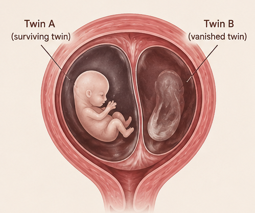
Vanishing Twin Syndrome, sometimes called disappearing twin syndrome, describes a kind of pregnancy loss. It happens when one embryo or fetus stops growing and gets reabsorbed into the body. This isn't an instant change, but rather a slow process. By the time of the next ultrasound, there might be nothing left to see.
There are different ways this can happen, depending mainly on when the loss occurs and whether the twins share a placenta.
In early resorption, which usually happens in the first trimester, the body either absorbs the embryonic tissue entirely, or the surviving twin's placenta does it. So, no trace of the dead twin is around by the next check-up. This is the most common way vanishing twins happen.
Another case is fetus papyraceus. When a twin doesn't grow in the second trimester, its tissue loses fluid and gets pressed against the growing twin and surrounding membranes. What's left looks thin, almost like paper, and could show up in the placenta at birth.
In rare cases, especially in monochorionic (shared placenta) pregnancies, some fetal material may be incorporated into the surviving twin's tissue.
Both involve pregnancy loss, but they are not the same thing. In a miscarriage, the pregnancy as a whole ends, and the tissue leaves the body through the cervix. In vanishing twin syndrome, only one baby is lost while the other continues to grow. The lost twin is reabsorbed rather than expelled. There may be light bleeding, but the overall pregnancy continues. This distinction matters medically, emotionally, and in terms of how the pregnancy is managed going forward.
The large majority of cases are identified in the first trimester, typically between six and twelve weeks. Early ultrasounds now routinely detect multiple gestational sacs or embryos, only for one to be absent on a scan a few weeks later.
Vanishing twin syndrome is more prevalent than previously thought, largely because early ultrasound technology is now so much more sensitive. Studies suggest it occurs somewhere between 20% and 30% of multiple pregnancies. In pregnancies conceived through IVF, where multiple embryos are often transferred, the rates can be even higher.

The exact reason one twin stops developing is not always identified, but several vanishing twin syndrome causes are well recognised.
This is the most common underlying cause. When an embryo carries chromosomal errors, extra chromosomes, missing chromosomes, or structural abnormalities, it cannot develop normally.
The pregnancy essentially self-selects: the embryo that is not viable stops growing while the healthy one continues.
A properly formed placenta is essential for fetal growth. When the placenta develops abnormally, whether due to poor implantation, structural defects, or inadequate blood vessel formation, it may not be able to sustain two growing babies. One twin receives insufficient support and stops developing.
For a pregnancy to progress, the fertilised egg must implant correctly in the uterine lining. When implantation is shallow or occurs in an unfavourable location, the embryo may lack the hormonal and nutritional support it needs.
The umbilical cord is the lifeline between the placenta and the fetus. Abnormalities in cord structure or insertion can reduce blood flow to one twin, compromising development.
In twin pregnancies where cords are poorly positioned or structurally abnormal, one fetus may not receive adequate oxygen and nutrients to survive.
Certain factors increase the likelihood of vanishing twin syndrome, though they do not directly cause it.
Advanced maternal age is associated with higher rates of chromosomal abnormalities in embryos. IVF conception, which often involves multiple embryos, naturally increases the chance of a multiple pregnancy and therefore of one embryo failing. Multiple gestation pregnancies also carry inherently higher rates of complications, including fetal loss.

Vanishing twin syndrome symptoms may be accompanied by the following signs and changes:
In a significant number of cases, there are no symptoms at all. The loss is discovered only when an ultrasound that previously showed two embryos no longer shows one.
Light vaginal bleeding during the first trimester is one of the more commonly reported vanishing twin syndrome symptoms. It is not always heavy or associated with pain, and many women mistake it for implantation bleeding or normal first-trimester spotting.
Some women experience mild cramping that feels similar to early miscarriage symptoms or menstrual discomfort.
Moderate or persistent pelvic pain, particularly if it accompanies bleeding, should always be evaluated by a healthcare provider.
Human chorionic gonadotropin (hCG) levels rise steadily in healthy pregnancies. When one twin is lost, hCG levels may plateau, rise more slowly than expected, or briefly dip before recovering.
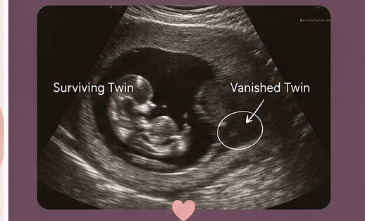
Diagnosis almost always begins with an early ultrasound, typically the six to eight-week scan that identifies two gestational sacs, two yolk sacs, or two embryos with heartbeats.
When a follow-up scan, often at ten to twelve weeks, shows only one embryo, and no alternative explanation exists (such as a scan error), the clinical picture points strongly to vanishing twin syndrome. The empty sac or absent embryo, or occasionally a faint remnant of tissue, confirms what happened.
Serial hCG measurements can help track whether the hormone is rising appropriately for a single pregnancy after the loss. A normal hCG trajectory following the event is generally reassuring.
When the loss occurs in the second trimester, the diagnosis may not be made until delivery. Examination of the placenta may reveal fetus papyraceus, the compressed remains of the second twin or other signs of a prior twin pregnancy. In these cases, parents may not know that vanishing twin syndrome occurred until after the baby is born.
The vanishing twin effect on the surviving twin largely depends on when the loss occurs.
When a twin is lost during the first trimester, which is the most common case, the outlook is generally excellent, and the remaining baby typically continues to develop normally with little to no increased risk of long-term health problems. However, losses occurring in the second or third trimester can be associated with a higher risk of complications, including preterm birth, growth restriction, and, in some cases, neurological injury.
The risk is greatest in monochorionic (shared-placenta) twins, where shared blood vessel connections can lead to sudden changes in blood flow after the loss of one twin. While many surviving babies are born healthy and achieve normal developmental milestones, pregnancies involving later losses or shared placentas usually require closer monitoring by a maternal-fetal medicine specialist.
Non-invasive prenatal testing (NIPT) analyses cell-free fetal DNA circulating in the mother's blood. After a twin is lost, residual DNA from that twin can remain in the maternal bloodstream for some time. This means NIPT may be reading DNA from two different individuals rather than one.
The presence of chromosomally abnormal DNA from the vanished twin can produce false-positive NIPT results for the surviving healthy baby.
There is no treatment for the vanished twin itself. Once an embryo has stopped developing and begun to be reabsorbed, nothing can reverse that process. Management focuses entirely on supporting the surviving baby and the mother.
Getting regular ultrasound checks to monitor how the baby's growing, how the placenta’s working, and overall well-being, too, is essential.

If you see vaginal bleeding or just spotting during your pregnancy, call your doctor right away. Also reach out to a medical professional if you feel more than mild pelvic pain or notice your pregnancy symptoms drop suddenly, or if anything worries you after a scan shows only one baby survived.
At Cloudnine, pregnancies affected by vanishing twin syndrome are supported through a coordinated approach involving fetal medicine specialists, obstetricians, radiologists, and neonatal experts. Advanced ultrasound imaging enables early and accurate diagnosis, while regular follow-up scans help monitor the health and growth of the surviving baby throughout the pregnancy.
Vanishing twin syndrome is both emotional, yet the surviving baby usually keeps developing normally, and complications are rare. Early diagnosis, regular check-ups, and advice from experts are key for a smooth outcome. Worried about a twin pregnancy? Reaching out to a specialist offers clarity, comfort, and the support you need through the journey.

Want to consult the best gynecologists in India? Please find the links below.
Want to consult the best Maternity Packages in India? Please find the links below.

A baby who was part of a twin pregnancy but whose co-twin did not survive is sometimes called as a twinless twin.
Most cases of vanishing twin syndrome occur in the first trimester, typically between six and twelve weeks of pregnancy.
No. Vanishing twin syndrome is actually quite common, occurring in an estimated 20–30% of multiple pregnancies. Because many cases happen before a first ultrasound is performed, the true incidence is likely higher than recorded statistics suggest.
No. Once an embryo has stopped developing, it cannot recover or resume growth. The tissue is gradually reabsorbed by the body. What is sometimes mistaken for a "return" is simply the detection of residual tissue on a later scan, which eventually disappears entirely.


