


When a baby is born prematurely, the focus in the days and weeks that follow is rightly on breathing, feeding, weight, and keeping the baby stable. Eye health is rarely the first thing on a parent's mind. Yet for preterm babies, a condition called retinopathy of prematurity (ROP) is one of the most important things to watch for and one of the most preventable causes of blindness in children in India.
Retinopathy of prematurity can develop silently, with no outward signs in the early stages. By the time it is visible to parents, it may have already progressed to a point where treatment is less effective, or the damage is irreversible. This is why ROP screening is not optional; it is essential for every baby born at risk.
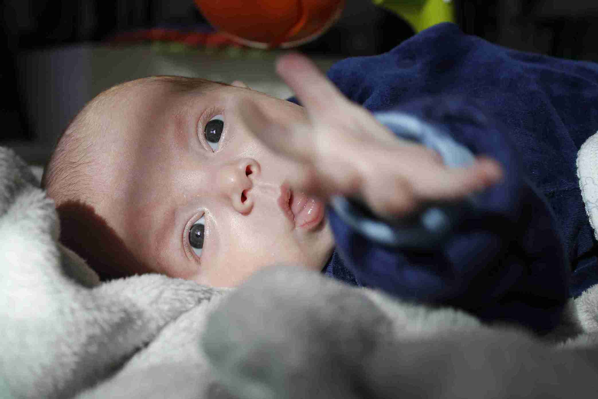
The retina is the tissue layer at the back of the eye. It detects light and sends visual signals to the brain. In a full-term baby, the blood vessels that supply the retina are fully developed by birth. In a preterm baby, this development is incomplete.
After a premature birth, the retinal blood vessels must continue to grow outside the womb, in an environment quite different from the protective, oxygen-controlled environment of the uterus. In some preterm babies, this process goes wrong. Instead of growing normally, the blood vessels grow abnormally, in a disorganised way that can cause scar tissue to form on the retina.
This scar tissue can pull on the retina and, in severe cases, cause it to detach from the back of the eye, leading to permanent vision loss or blindness.
Two main factors drive ROP in premature babies:
Oxygen exposure: The retinal blood vessels are sensitive to oxygen levels. In the NICU, premature babies often receive supplemental oxygen to support their breathing. High oxygen levels can temporarily stop the developing retinal blood vessels from growing. When oxygen is later reduced, the vessels try to grow quickly to compensate, but this rapid, disorganised growth causes the abnormal vessel proliferation of ROP.
Modern NICUs carefully monitor oxygen levels to minimise this risk, but it cannot be eliminated entirely, especially in very premature babies whose retinal development is almost incomplete at birth.
Degree of prematurity: The earlier a baby is born, the less developed the retinal blood vessels and the higher the risk of ROP. A baby born at 24 weeks has far more developing blood vessels to grow outside the womb than a baby born at 32 weeks.
The Indian Academy of Paediatrics recommends ROP screening for:
All babies born before 34 weeks of gestation
All babies born weighing less than 1.7 Kg
Babies between 34 and 36 weeks who had a significant illness or received supplemental oxygen
Additional risk factors that increase ROP in premature babies include blood transfusions, severe anaemia, infection, respiratory distress syndrome, and prolonged ventilation.
This is why routine screening is so critical. In the early stages, retinopathy of prematurity symptoms are not visible to parents or even to a general examination; the abnormal blood vessel growth happens at the back of the eye, beyond what is visible without specialist equipment.
By the time ROP produces symptoms that a parent might notice, a white reflex in the eye (leukocoria), crossed eyes, nystagmus (involuntary eye movements), or the baby not tracking light, the condition has usually reached a severe stage that is much harder to treat.
This is exactly why waiting for symptoms before seeking screening is dangerous. The goal of ROP screening is to identify the condition before symptoms appear, when treatment is most effective.
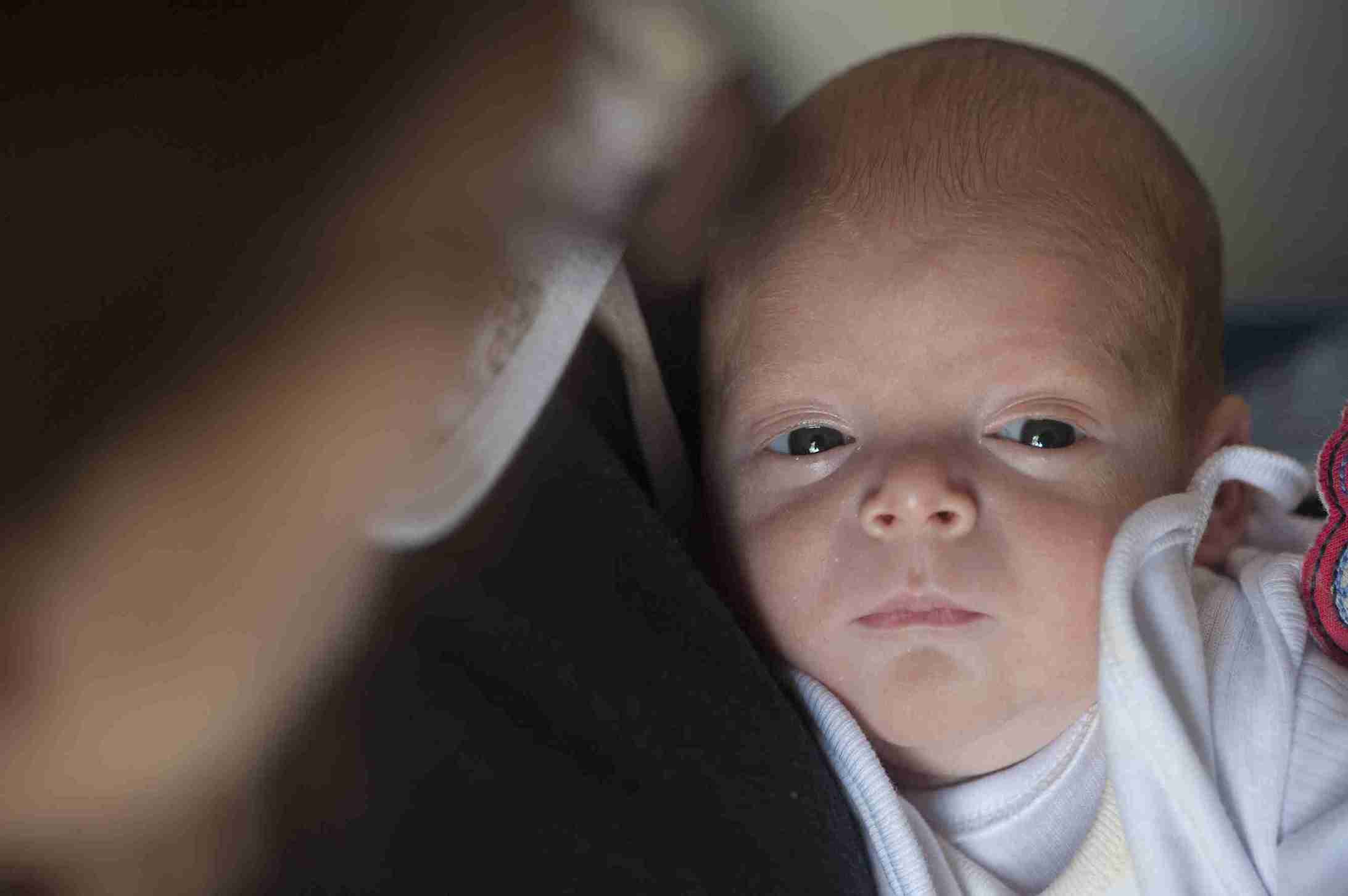
ROP screening for premature babies is done by an ophthalmologist with specialist training in paediatric retinal examination. It involves:
Dilating the baby’s pupils using eye drops
Examining the retina using an indirect ophthalmoscope
Documenting the stage (1, 2, 3 or 4) and location
ROP screening timing is based on the baby's gestational age and postnatal age. After the first examination, follow-up frequency depends on the findings. Mild changes may need weekly or biweekly review. Babies with no or minimal changes may be followed less frequently. The ophthalmologist will recommend a schedule based on the findings.
Most cases of ROP (Stages 1 and 2) resolve on their own without treatment. Regular monitoring is all that is needed. Treatment becomes necessary when ROP reaches what is called threshold or pre-threshold disease - Stage 3 with specific features - or when higher stages are reached.
Laser photocoagulation is the standard treatment. A laser destroys the peripheral retinal tissue where abnormal blood vessels are growing, stopping their abnormal proliferation and protecting the central retina where vision is focused. The procedure is done under sedation or general anaesthesia and is highly effective when done at the right time.
Anti-VEGF injections that block the growth factor driving abnormal vessel growth are increasingly used alongside or instead of laser therapy, especially in certain patterns of severe ROP. They are injected directly into the eye under local anaesthesia and have shown good results.
Surgery for advanced ROP (Stage 4 and 5) involving retinal detachment requires surgical intervention. At this stage, outcomes are less predictable, and vision recovery is limited, reinforcing why early detection through screening is so important.
India has a significant burden of ROP-related blindness, which is due to the increasing survival of very premature babies in NICUs and limited access to timely ROP screening.
Babies who are born in a hospital with a good NICU but do not have access to a trained ophthalmologist for screening are at significant risk of preventable blindness. Parents of premature babies should specifically ask, before discharge from the NICU, whether ROP screening has been done and whether follow-up screening appointments have been scheduled.
If a baby is transferred between hospitals, transfer ROP screening records with the baby's medical notes. Gaps in follow-up are one of the most common reasons severe ROP goes untreated until it is too late.

Retinopathy of prematurity is preventable in most cases, not prevented from occurring, but prevented from causing permanent vision loss, through timely screening and treatment. Every premature baby at risk deserves an eye examination at the right time, by a trained ophthalmologist, with appropriate follow-up. If your baby was born early, make sure ROP screening is part of the care plan before you leave the hospital.
Want to consult the best pediatrician in India? Please find the links below

ROP is a condition in which the blood vessels supplying the retina develop abnormally in premature babies. Because retinal vessel development is incomplete at the time of premature birth, the vessels grow in a disorganised way after birth, sometimes causing scar tissue that can pull the retina away from the back of the eye. In severe cases, this leads to permanent vision loss or blindness.
ROP screening for premature babies is essential because the condition causes no visible symptoms in its early stages; by the time a parent notices anything wrong, the disease may already be severe. Screening by a trained ophthalmologist identifies abnormal vessel growth before it reaches the stage of causing retinal damage, allowing treatment to be given at the point when it is most effective. Without screening, ROP can progress silently to permanent blindness.
ROP in premature babies is most likely in babies born before 34 weeks of gestation, babies weighing less than 1.7 Kgs at birth, and babies between 34 and 36 weeks who received supplemental oxygen or had a significant illness. In India, the Indian Academy of Paediatrics recommends broader screening criteria than in some other countries, reflecting local risk factors. Additional risks include blood transfusions, severe infection, and prolonged ventilation in the NICU.
The timing depends on gestational age at birth. In India, the first ROP screening is typically done at 4 weeks of postnatal age for babies born before 28 weeks, and at 31 weeks of corrected gestational age or 4 weeks of postnatal age, whichever is later, for babies born between 28 and 34 weeks. After the first exam, follow-up frequency depends on findings. Parents should confirm that screening is scheduled before the baby is discharged from the NICU.


 Best Gynecologists in Bellandur Best Gynecologists in Electronic City Best Gynecologists in HRBR LayoutBest Gynecologists in Jayanagar Best Gynecologists in Kanakapura RoadBest Gynecologists in Malleswaram Best Gynecologists in Nagarbhavi Best Gynecologists in Old Airport RoadBest Gynecologists in Sahakarnagar Best Gynecologists in Sarjapur RoadBest Gynecologists in ThanisandraBest Gynecologists in Varthur RoadBest Gynecologists in Whitefield Best Gynecologists in Industrial Area Phase IIBest Gynecologists in OMRBest Gynecologists in T NagarBest Gynecologists in New Industrial TownBest Gynecologists in IndirapuramBest Gynecologists in Golf Course RoadBest Gynecologists in Sector 14Best Gynecologists in Sector 47Best Gynecologists in Banjara HillsBest Gynecologists in GachibowliBest Gynecologists in Hitech CityBest Gynecologists in KompallyBest Gynecologists in Jawahar NagarBest Gynecologists in Gomti NagarBest Gynecologists in Phase 3, Sector 58Best Gynecologists in MaladBest Gynecologists in NerulBest Gynecologists in VashiBest Gynecologists in Kailash ColonyBest Gynecologists in PatparganjBest Gynecologists in Punjabi BaghBest Gynecologists in RohiniBest Gynecologists in VikaspuriBest Gynecologists in Gaur CityBest Gynecologists in Pari ChowkBest Gynecologists in Sector 51Best Gynecologists in Sector 5 Swastik ViharBest Gynecologists in Kalyani NagarBest Gynecologists in SB RoadBest Gynecologists in Pimple SaudagarBest Pediatricians in BellandurBest Pediatricians in Electronic CityBest Pediatricians in HRBR LayoutBest Pediatricians in JayanagarBest Pediatricians in Kanakapura RoadBest Pediatricians in MalleswaramBest Pediatricians in NagarbhaviBest Pediatricians in Old Airport RoadBest Pediatricians in SahakarnagarBest Pediatricians in Sarjapur RoadBest Pediatricians in ThanisandraBest Pediatricians in Varthur RoadBest Pediatricians in WhitefieldBest Pediatricians in Industrial Area Phase IIBest Pediatricians in OMRBest Pediatricians in T NagarBest Pediatricians in New Industrial TownshipBest Pediatricians in IndirapuramBest Pediatricians in Golf Course RoadBest Pediatricians in Sector 14Best Pediatricians in Sector 47Best Pediatricians in Hitech CityBest Pediatricians in Banjara HillsBest Pediatricians in GachibowliBest Pediatricians in KompallyBest Pediatricians in Jawahar NagarBest Pediatricians in Gomti NagarBest Pediatricians in Phase 3, Sector 58Best Pediatricians in MaladBest Pediatricians in NerulBest Pediatricians in VashiBest Pediatricians in Kailash ColonyBest Pediatricians in PatparganjBest Pediatricians in Punjabi BaghBest Pediatricians in RohiniBest Pediatricians in VikaspuriBest Pediatricians in Gaur CityBest Pediatricians in Pari ChowkBest Pediatricians in Sector 51Best Pediatricians in Sector 5 Swastik ViharBest Pediatricians in Kalyani NagarBest Pediatricians in SB RoadBest Pediatricians in Pimple Saudagar


